
S3 lands just after S2, an early diastolic thud from blood rushing into the ventricle. S4 lands just before S1, a late diastolic sound from the atria squeezing against a ventricle that will not stretch. That timing difference is the whole exam question, and it is also the fastest way to build a differential at the bedside.
Here is the rest of what you need before rounds or a shelf exam:
- S3 = ventricular (protodiastolic) gallop. Can be normal in a healthy 22-year-old; in a 68-year-old with leg swelling, it is a red flag for heart failure.
- S4 = atrial (presystolic) gallop. Almost never normal in adults. It usually points to a ventricle stiffened by hypertension, hypertrophic cardiomyopathy, or ischemia.
- Both are low-frequency sounds you will miss with the diaphragm. Use the bell.
Everything below builds on that one distinction: mechanism, mnemonics, causes, technique, and how to actually retain it under exam pressure.
Key Takeaways
S3 and S4 differ by timing and mechanism: S3 follows S2 from rapid filling into a compliant or overloaded ventricle, while S4 precedes S1 from atrial contraction against a stiff one.
| Point | Details |
|---|---|
| Timing rule | S3 comes right after S2 (early diastole); S4 comes right before S1 (late diastole). |
| Mnemonic pairing | Kentucky (S1-S2-S3) for S3; Tennessee (S4-S1-S2) for S4. |
| Age changes meaning | S3 can be normal in youth and pregnancy; S4 is almost always pathologic in adults. |
| Technique that works | Use the stethoscope bell, light pressure, and left lateral decubitus at the apex. |
| Confirm with timing | Correlate the sound against the radial pulse or ECG to resolve ambiguous cases. |
This article is general information, not a substitute for advice from a qualified doctor. Consult a qualified healthcare professional about your own circumstances before acting on anything here.
Table of Contents
- S3 vs S4 Timing and the Kentucky/Tennessee Mnemonics
- What Causes S3 and S4: The Physiology Behind Each Sound
- Clinical Significance: When S3 and S4 Signal Real Disease
- How to Hear S3 and S4 at the Bedside
- Turning Recognition Into Exam Points
- How Common Are S3 and S4, and Who Gets Them?
- Why S3 and S4 Matter for Prognosis and Management
- How S3 and S4 Show Up on Phonocardiography and Echocardiography
- Why S3 and S4 Are Genuinely Hard to Auscultate
- Telling S3 and S4 Apart From Other Extra Sounds
- Get Better at Recognizing These Findings, Faster
- Editorial Take: S3 vs S4 for Med Students
- Sources
- FAQ
S3 vs S4 Timing and the Kentucky/Tennessee Mnemonics
The four heart sounds run in a fixed sequence: S1 (mitral and tricuspid closure), systole, S2 (aortic and pulmonic closure), then diastole, then back to S1. S3 and S4 are the two spots where an extra sound can slot into that diastolic gap, and they occupy opposite ends of it. S3 shows up in early diastole, during the rapid, passive filling phase right after the mitral valve opens. S4 shows up in late diastole, during atrial contraction, the "atrial kick" that tops off ventricular filling right before the next S1.
The mnemonics work because they mimic the actual rhythm of the syllables:
- Kentucky for S3: say it out loud and the stress falls "Ken.TUCK.y," matching S1-S2-S3, with S3 trailing softly after the second heart sound.
- Tennessee for S4: "TEN.nes.see" front-loads the stress, matching S4-S1-S2, with S4 arriving just ahead of the first heart sound.
- Practice both mnemonics against a real audio recording before you try them on a patient. Your ear needs the pattern locked in before you can pull it out of a noisy room.
The pitfall that trips up most third-year students is not S3 versus S4. It's mistaking a widely split S2 or an early systolic click for a diastolic gallop. A split S2 occurs at the very end of systole, not in diastole, and it moves with respiration. A gallop does not. If you are unsure whether a sound is S3, S4, or a split S2, time it against the carotid pulse: S3 and S4 both fall in diastole, well after the carotid upstroke, while a split S2 sits right at the upstroke's end.
Pro Tip: Hum "Kentucky" and "Tennessee" under your breath while you auscultate a standardized patient. It sounds ridiculous out loud, but timing the syllables to what you're hearing is faster than trying to count milliseconds in your head during an OSCE.
What Causes S3 and S4: The Physiology Behind Each Sound
S3 and S4 are not caused by valve clicks or turbulent flow the way murmurs are. They are low-frequency vibrations generated by the ventricular wall and the blood column itself, and the mechanism behind each one tells you almost everything about when it is dangerous.
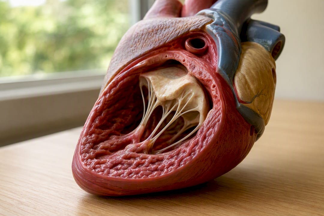
S3 comes from rapid filling meeting a ventricle that cannot smoothly absorb it. Right after the mitral valve opens, blood surges into the ventricle during the rapid filling phase. In a young, compliant, easily stretched ventricle, this happens quietly. In a ventricle that is either overloaded with volume (as in heart failure) or dilated (as in dilated cardiomyopathy), that inflow decelerates abruptly against the ventricular wall, and that sudden deceleration is what you hear as S3.
S4 comes from a stiff ventricle fighting back against atrial contraction. Late in diastole, the atria contract to deliver the final push of blood into the ventricle, the atrial kick. In a normal, compliant ventricle, this transition is silent. In a ventricle stiffened by long-standing hypertension, aortic stenosis, or hypertrophic cardiomyopathy, that same atrial push generates a palpable, audible vibration because the muscle wall resists the added volume.
Age changes the calculus for both sounds:
- A physiologic S3 is common in children, adolescents, and trained athletes because their ventricles are highly compliant and their stroke volumes run high.
- An S4 in a healthy 20-year-old is unusual enough that it should prompt a closer look, even without symptoms.
- Past age 40, a new S3 shifts the working diagnosis toward volume overload far more often than it does in a 19-year-old with a low resting heart rate.
One detail that catches students off guard on rounds: S4 is described as almost always pathologic in adults, a stronger statement than what's said about S3. That asymmetry is worth memorizing on its own, because it changes how urgently you should chase a workup.
Clinical Significance: When S3 and S4 Signal Real Disease
Whether either sound worries you depends almost entirely on the patient standing in front of you. A 24-year-old marathon runner with an S3 and a 70-year-old with peripheral edema and an S3 are not the same clinical picture, even though the sound is identical on the recording.
S3 shows up with these common causes:
- Heart failure with volume overload, especially decompensated systolic failure
- Dilated cardiomyopathy
- Significant mitral regurgitation, from the extra volume flooding the ventricle each beat
- Physiologic states: children, adolescents, pregnancy, and endurance athletes, where a compliant ventricle simply handles a bigger stroke volume without strain
S4 shows up with these common causes:
- Long-standing, poorly controlled hypertension, the single most common cause you'll see on the wards
- Left ventricular hypertrophy from any cause
- Hypertrophic cardiomyopathy, where a young patient with an S4 and a systolic murmur should raise real concern
- Aortic stenosis, from the pressure-overloaded ventricle stiffening over time
- Acute ischemia, where a new S4 can appear transiently during an anginal episode
Three quick vignettes show how this plays out on an exam stem:
A 74-year-old with a history of myocardial infarction presents with dyspnea, bilateral pitting edema, and jugular venous distention. Auscultation reveals a soft sound just after S2 at the apex. The combination points to systolic heart failure with an S3, not S4, because the extra volume and dilated chamber create the classic ventricular gallop pattern described by the American Heart Association.
A 55-year-old with a decade of uncontrolled blood pressure and no edema presents for a routine physical. A sound just before S1 at the apex, with a sustained, laterally displaced apical impulse, points toward LVH and an S4 rather than any volume-overload process. A 16-year-old athlete with no symptoms and a soft early diastolic sound that disappears when she stands up is very likely a benign physiologic S3, not a diagnosis in progress.
How to Hear S3 and S4 at the Bedside
Technique separates the students who can identify these sounds reliably from the ones who only catch them by accident.
- Use the bell, not the diaphragm. S3 and S4 are low-frequency sounds, and the bell transmits low frequencies far better than the diaphragm, which is built for high-frequency murmurs. Apply only light pressure, since pressing the bell firmly against the skin turns it into a diaphragm and filters out the very sound you're chasing.
- Position the patient in left lateral decubitus and place the bell at the cardiac apex, roughly the fifth intercostal space at the midclavicular line. This tips the heart closer to the chest wall and is the single highest-yield position for both gallops, according to PhysDx's auscultation technique guide. Ask the patient to exhale and briefly hold, since a breath-holding pause clears out lung sounds that mask a quiet gallop.
- Time the sound against the radial pulse or a single-lead ECG strip. S3 follows the pulse upstroke; S4 precedes it. This single habit eliminates most of the "wait, was that S3 or S4" confusion that trips students up on practical exams.
Maneuvers can help you confirm what you're hearing. Isometric handgrip raises afterload and can accentuate an S4 in a stiff ventricle. Squatting increases venous return and preload, which can bring out a subtle S3. Valsalva, by contrast, tends to decrease both sounds during the strain phase, useful mainly for distinguishing gallops from murmurs that behave differently under the same maneuver.
Pro Tip: If you genuinely cannot decide whether you're hearing S3 or S4, check the patient's pulse with your free hand while you listen. A sound that lands right where your finger feels the pulse is almost certainly S4; a sound that trails just after it is almost certainly S3.
Turning Recognition Into Exam Points
Recognizing a gallop on a real patient is one skill. Picking the right answer on a vignette-based question is a slightly different one, and it rewards a narrower set of memorized facts.
Lock in these exact associations before test day:
- S3 timing phrase: "early diastole, right after S2"
- S4 timing phrase: "late diastole, right before S1"
- S3 default suspicion in a patient over 40: volume overload or systolic heart failure
- S4 default suspicion in any adult: decreased ventricular compliance, most often from hypertension or LVH
- Auscultation site and tool: apex, bell, light pressure, left lateral decubitus
A study plan that works for this topic looks less like rereading a textbook chapter and more like structured, repeated exposure. Supervised bedside rounds where an attending confirms what you're hearing build the ear faster than any recording alone. Audio libraries of real heart sounds, paired with spaced-repetition flashcards that force you to name the sound before you see the answer, cement the timing. Question blocks written in the same stem style you'll actually face, USMLE-style stems that bury the S3/S4 clue inside a longer clinical vignette, train you to extract the relevant detail under time pressure rather than in isolation.
That last piece is where a lot of otherwise solid students lose points, not because they don't know the physiology, but because they never practiced pulling it out of a paragraph full of distractors. BoardMaster builds practice questions directly from uploaded lecture notes, so the cardiology set you study reflects the exact emphasis your professor put on S3 versus S4, rather than a generic bank's version of the topic.
How Common Are S3 and S4, and Who Gets Them?
Age is the single biggest driver of whether either sound counts as normal. A physiologic S3 is common enough in children and young adults that many clinicians consider it an expected finding on a routine pediatric exam, tied to the naturally high compliance and brisk filling of a young ventricle. That same sound in a patient past midlife carries far more weight, since ventricular compliance declines with age and a "gallop" at 65 usually means something the heart is compensating for rather than a benign variant.
Pregnancy is another physiologic S3 setting worth knowing cold, since the expanded blood volume and higher cardiac output of pregnancy can produce an audible S3 in an otherwise healthy patient. Trained endurance athletes fall into the same bucket, with resting bradycardia and elevated stroke volume creating filling dynamics that mimic what a diseased heart does under different circumstances.
S4 skews the opposite direction demographically. It becomes more common with age simply because the conditions that cause it, chronic hypertension, aortic stenosis, and age-related ventricular stiffening, accumulate over decades. A teenager with an S4 is a genuine outlier and warrants a closer look for hypertrophic cardiomyopathy. A 60-year-old with an S4 and a long blood pressure history is closer to an expected finding than a surprising one. Neither sound has a dramatic sex-based prevalence difference worth memorizing separately from the underlying disease patterns that drive them.
Why S3 and S4 Matter for Prognosis and Management
An S3 in a patient with known systolic heart failure is not just a diagnostic curiosity. It correlates with elevated filling pressures and worse functional status, and its presence has been tied to higher risk in heart failure populations, part of why the American Heart Association treats gallop detection as a meaningful data point in ongoing management, not just an admission-day finding. A new S3 that appears during a hospital stay, or one that resolves after diuresis, gives you a low-cost, real-time signal about whether treatment is actually reducing volume overload.
S4 carries a different kind of prognostic weight. Because it reflects chronic structural change, a stiffened, hypertrophied ventricle, it points less toward an acute crisis and more toward the downstream risk of that structural change: arrhythmia, diastolic dysfunction, and eventual heart failure with preserved ejection fraction if the underlying hypertension or outflow obstruction goes uncontrolled.
Neither sound stands alone in a management decision. Both should push you toward the next diagnostic step rather than serve as the final answer, and both belong in the same sentence as blood pressure control, volume status assessment, and imaging, not as an isolated exam pearl.
How S3 and S4 Show Up on Phonocardiography and Echocardiography
Auscultation is subjective, which is exactly why objective tools exist to confirm what your ear picks up. Phonocardiography records the actual sound waveform and timestamps it against the ECG, making it the closest thing to ground truth for timing questions. On a phonocardiogram, S3 appears as a low-frequency deflection shortly after the T wave, while S4 appears as a low-frequency deflection just before the QRS complex, a visual version of the same "before S1, after S2" rule you use at the bedside.
Echocardiography does not directly detect the sound, but it confirms the mechanism behind it. A patient with an auscultated S3 and a dilated, poorly contracting left ventricle on echo has a coherent picture: reduced ejection fraction correlating with the gallop you heard. A patient with an S4 and echo findings of increased wall thickness or diastolic dysfunction similarly ties the sound to its structural cause. When the auscultation finding and the echo findings disagree, or when a sound is genuinely ambiguous, correlating with echocardiography rather than trusting the ear alone is the responsible next step, and it's a pattern worth internalizing well before residency.
Why S3 and S4 Are Genuinely Hard to Auscultate
Even experienced clinicians disagree on gallops, and there are real reasons why. Both sounds are low-frequency and low-amplitude, sitting right at the edge of human hearing, which means ambient noise, a thick chest wall, obesity, or lung hyperinflation from COPD can bury them entirely. A tachycardic patient compresses diastole into a shorter window, making it harder to separate a true S3 or S4 from a rapid, closely spaced S1 and S2.
Interobserver reliability is a known weak point in cardiac auscultation generally, and gallops are among the sounds clinicians disagree on most. That is not a reason to skip the skill. It is a reason to treat a single auscultation finding as one data point rather than a definitive diagnosis, and to lean on positioning, the bell, and pulse timing rather than assuming your ear alone is infallible on the first pass. It's also why structured bedside supervision matters more here than for most physical exam findings. A skill this dependent on subtle acoustic discrimination improves with repetition and correction far more than it does with reading alone.
Telling S3 and S4 Apart From Other Extra Sounds
A few other findings get confused with gallops often enough to deserve a direct comparison. An opening snap, heard in mitral stenosis, occurs early in diastole like S3 but is higher pitched, sharper, and heard best with the diaphragm rather than the bell, a useful discriminator when the timing alone is ambiguous. A pericardial knock, seen in constrictive pericarditis, also lands in early diastole but tends to be louder and occurs slightly earlier than a typical S3, alongside other signs of constriction like Kussmaul's sign.
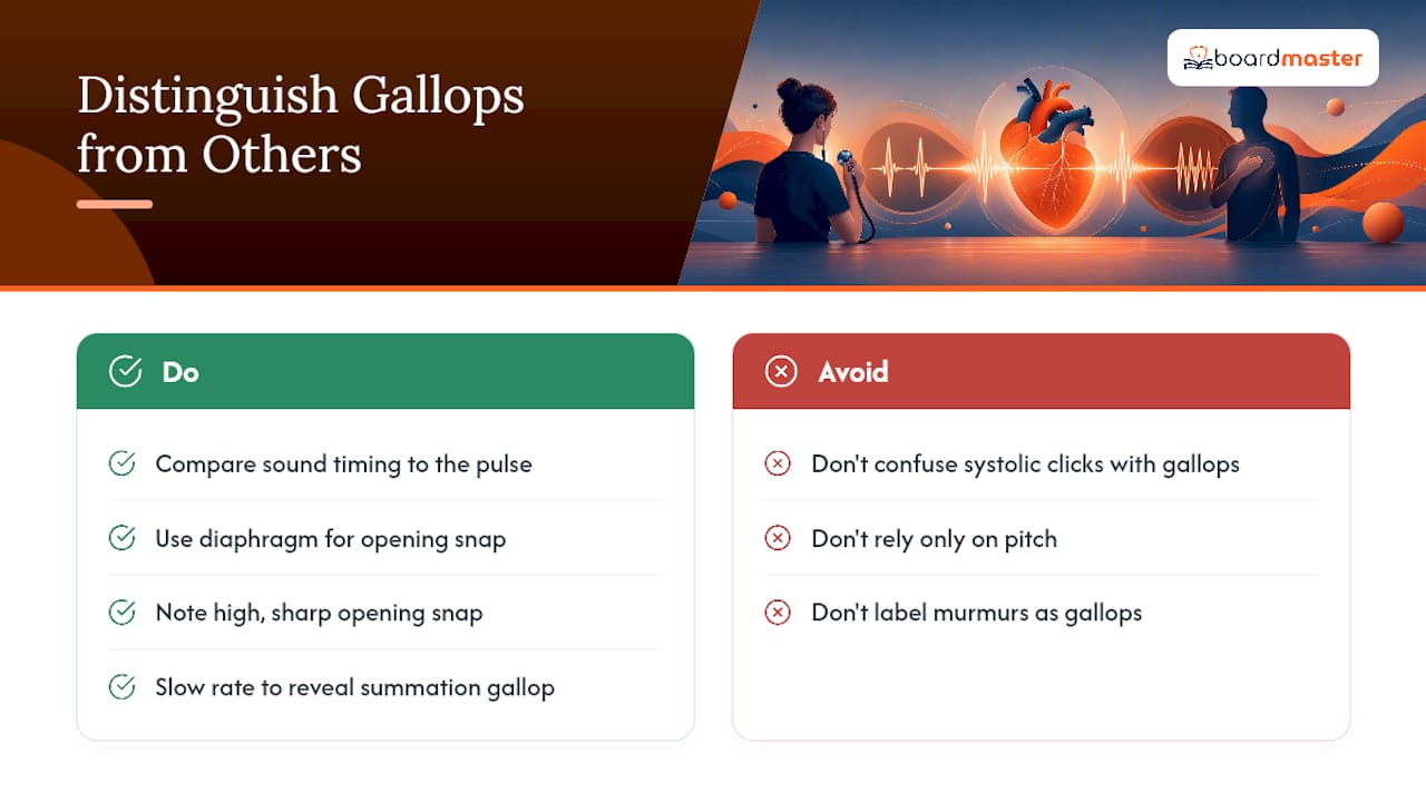
Systolic clicks, such as the mid-systolic click of mitral valve prolapse, sit in an entirely different phase of the cardiac cycle and should never be confused with a diastolic gallop if you're timing correctly against the pulse. A summation gallop, which happens when tachycardia compresses diastole enough that S3 and S4 fuse into a single sound, is its own trap: it can sound like one loud gallop when it is actually two superimposed ones, and slowing the heart rate (or catching the patient at a lower rate) sometimes unmasks the two components separately.
The differential also has to include murmurs, since a soft, low-pitched S3 or S4 can be mistaken for a decrescendo murmur by an inexperienced ear. The fix is the same one used throughout this guide: a true gallop is a single discrete sound with a fixed timing, while a murmur has duration and typically changes character across the phase of the cycle it occupies.
Get Better at Recognizing These Findings, Faster
The physiology and technique above will get you through most exam stems and most bedside encounters, but retention is the real bottleneck for this topic. Students tend to relearn S3 versus S4 from scratch every time it resurfaces on a shelf exam because the first pass never gets reinforced with enough targeted repetition.
BoardMaster's AI question generator builds USMLE-style stems directly from the lecture slides your cardiology professor actually used, so the gallop questions you practice reflect the specific emphasis and framing your course puts on the topic rather than a generic bank's version of it. Pair that with the platform's lean study workflow, which uses spaced repetition to resurface high-yield findings like S3 and S4 right before they'd otherwise fade, and you spend less time rereading and more time answering the exact style of question you'll see on test day. For students layering critical-care or nursing-track exam prep on top of core cardiology, the CCRN cardiovascular study guide covers the same auscultation fundamentals from a bedside-monitoring angle worth reviewing alongside your board prep.
Editorial Take: S3 vs S4 for Med Students
Most review material treats S3 and S4 as a memorization pair: two labels, two timings, done. That framing gets students through a quiz and fails them on the wards, because the harder skill isn't naming the sound. It's deciding how worried to be once you've heard it.
The conventional advice undersells age and context. A gallop chart that lists "S3: heart failure" without immediately pairing it against "S3 in a healthy 20-year-old: normal" sets students up to over-call benign findings or, worse, under-call a real one in an older patient because they never practiced the judgment call, only the label.
What actually moves the needle is repetition against real clinical framing, not more flashcard passes on isolated definitions. Practice the sound alongside the patient's age, volume status, and blood pressure history every single time, because that's the actual test you'll face on rounds and in most vignette-based exam questions. Technique matters too. Skipping the bell or the lateral decubitus position wastes a finding you'd otherwise catch cleanly. Prioritize pairing mechanism with context before you worry about polishing your stethoscope technique further.
— Dr. Ahmed Abuzoor
Sources
- NCBI Bookshelf: Third heart sound (S3) / ventricular gallop
- PhysDiX / PhysDx: Heart auscultation — additional sounds and technique
FAQ
What does an S3 and S4 heart sound indicate?
S3 indicates rapid ventricular filling and, in adults, often signals volume overload or heart failure; S4 indicates atrial contraction against a stiff, noncompliant ventricle, usually from hypertension or hypertrophy.
Is S3 Tennessee or Kentucky?
S3 is Kentucky, matching the S1-S2-S3 cadence. S4 is Tennessee, matching the S4-S1-S2 cadence.
What do S1, S2, S3, and S4 mean?
S1 and S2 mark valve closures (mitral/tricuspid and aortic/pulmonic), while S3 and S4 are extra diastolic sounds, a ventricular gallop from rapid filling and an atrial gallop from atrial contraction, respectively.
How do you tell S3 apart from S4 at the bedside?
Time the sound against the radial pulse or a single ECG lead: S3 follows the pulse upstroke, and S4 precedes it, using the stethoscope bell at the cardiac apex for both.