
High-yield images are the pictures that reliably cue a tested diagnosis or classic sign, the ones examiners return to year after year because they teach a concept fast. Your best first move is opening the AMSER "must-see" list on Radiopaedia and treating it as a checklist, then locking each case into memory with active recall and spaced repetition rather than passive scrolling.
TL;DR:
- Radiopaedia's AMSER collection provides the most relevant high-yield cases aligned with NBME-style exams, making it the top starting point for effective visual study.
- Mastering images involves active recall methods such as describing findings aloud and building multiple prompts, rather than passive recognition.
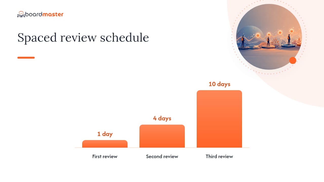
- Reviewing images on a schedule of 1, 4, and 10 days with interleaved modalities improves long-term retention and exam readiness.
- Classic findings like pneumothorax, hyperdense MCA sign, and bowel obstruction are the most frequently tested imageries across different body systems.
- Uploading lecture slides into platforms like BoardMaster personalizes high-yield practice, merging class and board exam preparation efficiently.
Table of Contents
- Where to Find High Yield Images for Medicine Right Now
- How to Study With Medical Images So They Actually Stick
- The Must-See Imaging Checklist by Body System
- A Repeatable Framework for Reading Exam Images
- How BoardMaster Turns Lecture Images Into High-Yield Practice
- Memorize the Classics, but Train the Framework
- Turn Your Own Lectures Into an Image Study Engine
- Sources
- FAQ
Where to Find High Yield Images for Medicine Right Now
Not every image bank deserves your limited study hours. Some are built for radiologists finishing fellowships; others are built for you, right now, three weeks before Step 2 CK.
Start with Radiopaedia's AMSER collection. It maps 26 classic cases directly onto the imaging findings NBME-style exams test most often, and every entry links to a full article plus practice questions. Treat it like a punch list: open one case, name the finding out loud, then check it off.
Beyond AMSER, here's what actually earns a spot in a tight rotation:
- NBME-style image PDFs and compact image guides. These condense findings into single-page visual summaries, ideal for a five-minute scan before bed rather than deep study.
- High-yield video playlists. Rapid-fire review videos train pattern recognition at exam speed, which matters more than most students realize until they're staring down a 60-second countdown.
- Flashcard decks (Quizlet, Anki-style). Vet these carefully. A deck with wrong labels or blurry scans teaches you the wrong reflex just as fast as a good one teaches you the right one.
- Histology and pathology image banks. Boards still test classic micro findings, and gross anatomy sets fill the gap that radiology alone will not cover.
- Institutional and open-education image sets. Prefer these, along with Radiopaedia's own library, when you need images you can legally reuse in your own study materials.
The ABR Essentials of Radiology guide organizes acute imaging topics by modality, which is a useful cross-check if your source material feels scattered.
How to Study With Medical Images So They Actually Stick
Staring at an image and thinking "yeah, I recognize that" is the single biggest trap in visual study. Recognition without recall is a false signal, and it evaporates the moment you're under exam pressure.
Use this sequence instead:
- Cover, label, describe, compare. Cover the caption, name the finding out loud, describe why it looks that way, then compare it against a lookalike differential.
- Build three prompt types per image. A diagnosis prompt ("What is this?"), a finding prompt ("Describe what you see"), and a differential prompt ("What else could cause this pattern?").
- Space your reviews. Revisit a new image at 1 day, 4 days, and 10 days out, folding it into whatever question bank rotation you already run.
- Interleave modalities. Mix a chest x-ray, a brain MRI, and a histology slide in the same session instead of blocking by system. Pattern-only learners fall apart when the order gets scrambled on exam day, and interleaving is the fix.
- Run timed rapid-fire rounds. Ten images, sixty seconds each, forces the recognition speed real exams demand.
- Mine your own lectures. Pull one or two professor-emphasized images per lecture and turn each into a focused card rather than transcribing the whole slide.
Pro Tip: Cap each image card at one testable idea. A card that tries to teach the finding, the mechanism, and three differentials at once is really three cards wearing a trench coat.
For a fuller framework on turning raw lecture slides into a sane daily rotation, see BoardMaster's lean study workflow guide.
The Must-See Imaging Checklist by Body System
Every board-style exam draws from a fairly predictable pool of classic findings. Here's the shortlist organized by system, with the visual anchor to memorize for each.
Thorax: pneumothorax (look for the visceral pleural line), lobar consolidation with air bronchograms, and pleural effusion layering on a decubitus film.
Neuro: the hyperdense MCA sign for early ischemic stroke, the empty delta sign in dural venous sinus thrombosis, the caput medusae pattern of a developmental venous anomaly, and the insular ribbon sign. Worth knowing: the empty delta sign shows up in only a minority of confirmed sinus thrombosis cases, which a pictorial review of 20 classic neuroradiology signs flags as a real limitation, not a rare exception.
MSK and trauma: lipohemarthrosis on a knee film after trauma, plus the handful of fracture patterns and locations that repeat across question banks.
Abdomen and pelvis: free intraperitoneal air under the diaphragm, and dilated bowel loops with air fluid levels signaling obstruction.
Pediatrics: normal myelination timelines on MRI and the age-appropriate variants that get misread as pathology by students trained only on adult scans.
Histology and pathology: the "fried egg" appearance of oligodendrocytes and drusen deposits in retinal pathology remain staples on board-style pathology sections.
A Repeatable Framework for Reading Exam Images
Every image vignette rewards the same sequence of questions, and running it consistently beats hoping the right answer jumps out at you.
Start with modality. Is it x-ray, CT, MRI, or ultrasound, and does the sequence or window matter? Radiologyinfo is a solid quick reference for how modality choice and patient prep change what you're looking at. Next, orient yourself: anterior or posterior, axial or coronal, T1 or T2. Then locate the anatomic landmarks before hunting for the abnormality; skipping this step is how students misname a normal variant as pathology.
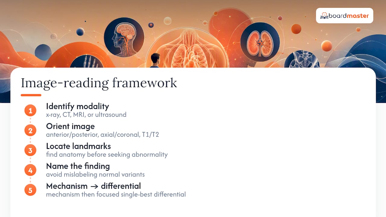
Once you've named the finding, ask what mechanism produces it, then narrow to the differential the exam is actually testing. Exam writers usually want the single-best-answer that fits urgency and classic presentation, not the rarest possible diagnosis.
Watch for two pitfalls. First, some classic signs have real sensitivity limits, so don't anchor on one sign's presence or absence. Second, resist the urge to over-rely on a memorized pattern when the vignette gives you clinical context that should redirect your differential entirely.
How BoardMaster Turns Lecture Images Into High-Yield Practice
Upload your lecture slides, and BoardMaster auto-generates image flashcards and USMLE-style image questions built around what your professor actually emphasized, then schedules them with spaced repetition. One student reported improved exam performance and efficiency while using this targeted approach.
A workable routine: convert one lecture set into a 10 to 15 minute daily image review. Dr. Ahmed Abuzoor's author profile covers more on aligning lecture emphasis with board-relevant content, and the common exam prep misconceptions guide walks through why memorization alone falls short.
Memorize the Classics, but Train the Framework
Speed comes from memorizing classic images cold. Judgment comes from a systematic read every time, not just when you're stuck. Keep your rotation lean, protect a rest day, and pick one interpretive habit, run it on every image you see, and never skip it.
— Dr. Ahmed Abuzoor
Turn Your Own Lectures Into an Image Study Engine
Generic question banks give you someone else's idea of what matters. BoardMaster gives you your professor's. Upload the slide deck your class actually uses, and the platform builds image flashcards and USMLE-style questions around the exact findings your instructor highlighted, then runs them through spaced repetition so nothing fades before exam day.

That matters because board exams and class exams don't always test the same emphasis, and studying only one set of material leaves gaps in the other. The platform merges preparation for board and class exams into a single study rotation instead of maintaining separate study tracks. Medical schools and student groups exploring this for a full cohort can check BoardMaster's partnership options to see how the workflow scales beyond one student's laptop. If you're studying solo, start by uploading your next lecture set and see what your first image deck looks like within minutes.
This article is general information, not a substitute for advice from a qualified doctor. Consult a qualified healthcare professional about your own circumstances before acting on anything here.
Sources
FAQ
What Does High Yield Mean in Medicine?
High yield refers to information, including images, that appears disproportionately often on exams relative to how much time it takes to learn, which is why a curated checklist like the AMSER list outperforms random image scrolling.
What Are Some High Yield Topics in Medicine?
Classic radiology signs (pneumothorax, hyperdense MCA sign, empty delta sign), common fracture patterns, obstructive bowel findings, and staple histology images like the oligodendrocyte "fried egg" appearance all repeat across board-style exams.
What Are the Most High Yield Topics for the USMLE Step 2 CK Exam?
Step 2 CK leans heavily on clinical image interpretation tied to management decisions, so chest and abdominal x-ray findings, stroke imaging signs, and OB/pediatric ultrasound basics tend to carry the most weight.
How Many Images Should I Actually Memorize?
There's no fixed number, but the AMSER list's 26 core cases represent a reasonable floor rather than a ceiling, and tools like BoardMaster can help you expand that set based on what your own lectures emphasize.
Are Video Playlists Better Than Flashcards for Image Review?
Neither replaces the other. Playlists build fast pattern recognition, while flashcards force active recall, and a strong rotation like BoardMaster's spaced-repetition decks uses both.